
Understanding Cranio-Cervical Instability
Cranio-cervical instability (CCI) involves abnormal movement where the skull meets the upper cervical spine. This abnormal movement can compromise the alignment and stability of the head and neck, leading to symptoms such as persistent headaches, neck pain, dizziness, and neurological issues. Causes of CCI include traumatic injuries, congenital or genetic disorders like Ehlers-Danlos syndrome, and degenerative changes associated with aging.
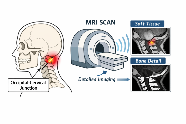
The symptoms of CCI often overlap with other conditions, which can make achieving an accurate diagnosis more difficult. Advanced imaging, particularly a cervical MRI scan, plays a critical role in identifying structural issues that may be contributing to a patient’s symptoms. This type of imaging provides detailed views of both soft tissues and bony structures in the cranio-cervical region, helping clinicians distinguish CCI from other disorders with similar presentations. Early identification of these abnormalities can guide treatment decisions and improve overall quality of life.
For individuals with suspected instability, symptoms may become more noticeable during certain head movements or physical activities. Because of this variability, a comprehensive imaging approach is important, as standard static images may not always capture subtle abnormalities responsible for discomfort or functional limitations.
Role of MRI in CCI Diagnosis
Magnetic Resonance Imaging (MRI) offers non-invasive, detailed visualization of the body’s internal structures. This technology uses strong magnetic fields and radio waves to create high-contrast images that highlight the intricate details of soft tissue, ligaments, intervertebral discs, and the spinal cord. In cranio-cervical instability, MRI is particularly valuable for identifying ligamentous injury or neural-structure damage that can be missed on standard radiographs or computed tomography (CT) scans.
One significant advantage of MRI is its absence of ionizing radiation, allowing for repeated assessments of individuals who require ongoing monitoring. Healthcare professionals rely on MRI’s clarity to evaluate the full spectrum of cranio-cervical anatomy, from the bony atlas and axis vertebrae to the supporting ligaments. This comprehensive view is crucial when planning conservative or surgical interventions.
Dynamic MRI Techniques
Conventional MRI scans are typically performed with the patient lying flat, providing a static view of tissues and bones. However, cranio-cervical instability can be dynamic, only appearing when the patient moves their neck or head into specific positions. Dynamic MRI techniques address this limitation by capturing images while the patient performs particular movements or is positioned upright. This method uncovers abnormalities that may be absent when the patient is still, bridging a critical gap in diagnosis.
Dynamic MRI has shown marked value, especially in individuals whose symptoms are provoked by particular head positions. For example, a systematic review highlighted how dynamic MRI consistently revealed motion-dependent stenosis and intramedullary signal changes not visible on static imaging. By using these specialized imaging sequences, doctors can better match radiographic findings to patient-reported symptoms and day-to-day function, thereby improving diagnostic accuracy and guiding personalized treatment strategies. ([pubmed.ncbi.nlm.nih.gov](https://pubmed.ncbi.nlm.nih.gov/41517513/))
Advantages of MRI Over Other Imaging Modalities
CT scans are superior for visualizing bony architecture but fall short of depicting the soft-tissue detail needed to assess CCI thoroughly. MRI delivers high-resolution images of ligaments, intervertebral discs, and the spinal cord. This capability is critical since ligament injuries are often central to cranio-cervical instability. Additionally, advances in MRI technology now allow for upright or weight-bearing imaging, which helps evaluate instability that only appears under physiological stress. The versatility of MRI, both in static and dynamic imaging, sets it apart from other modalities for a holistic assessment of the cranio-cervical junction.
Clinical Evidence Supporting MRI Use
The role of MRI in diagnosing CCI has been reinforced by a growing body of clinical research. For instance, a systematic review published in the Journal of Clinical Medicine highlighted how dynamic MRI consistently revealed motion-dependent stenosis and intramedullary signal changes not visible on static imaging. This method advances the potential for personalized treatment recommendations and early intervention. Additional studies have shown that MRI reliably detects injuries and instability that may otherwise be undetectable on X-ray or CT alone. A review on spinal cord injury by the American Association of Neurological Surgeons discusses how MRI outperforms other imaging methods for soft-tissue and neural visualization, emphasizing its critical role in neurosurgical and spinal assessment protocols.
Importance of Early Diagnosis
Detecting cranio-cervical instability early is key to preventing worsening symptoms and long-term neurologic complications. Early diagnosis allows healthcare professionals to initiate proper treatment strategies such as physical therapy, bracing, or surgical stabilization. When diagnosis is delayed, patients may suffer from chronic pain, functional decline, and potentially irreversible nerve injury. The use of high-quality, dynamic, and upright MRI imaging ensures that subtle instabilities do not go unnoticed, paving the way for prompt and effective care.
MRI’s unique ability to simultaneously visualize both bone and soft tissues under different conditions is foundational in the timely recognition and management of CCI. Patients benefit not only from the comprehensive scope of MRI but also its safety profile, supporting better long-term outcomes for those at risk of instability-related problems.
Conclusion
MRI scans are indispensable for diagnosing and managing cranio-cervical instability. Dynamic and upright MRI expand the physician’s diagnostic toolkit, enabling the visualization of position-dependent instabilities that static imaging could overlook. With its detailed images of soft tissues, ligaments, and the spinal cord, MRI supports more accurate diagnosis, effective treatment planning, and improved patient outcomes. Patients, caregivers, and clinicians should be aware of MRI’s role as the preferred imaging modality for patients with suspected cranio-cervical instability, enabling timely and appropriate interventions.